What is Retinal Vascular Occlusion?
Your retina is the light-sensitive layer at the back of your eye, and it depends on a steady blood supply to work. Blood arrives to it through the central retinal artery and drains away through the central retinal vein. Retinal vascular occlusion is what happens when one of these vessels or one of their smaller branches becomes blocked.
Both artery and vein occlusions can cause vision loss, although they differ in their underlying mechanisms and urgency. Retinal artery occlusions are less common but are ophthalmic emergencies because the retina can sustain irreversible damage rapidly when its blood supply is interrupted.
Retinal vein occlusions can also cause significant vision loss and complications such as macular oedema and abnormal blood vessel growth. They are generally less immediately urgent than artery occlusions, although they still require timely assessment and treatment.
Sudden, painless loss of vision in one eye should be treated as an emergency. Seek immediate medical attention rather than waiting for a scheduled appointment.
Types of Retinal Vascular Occlusion
Retinal vascular occlusions are broadly divided into retinal artery occlusions and retinal vein occlusions. Each can be further classified according to whether the central vessel or one of its branches is affected.
Artery occlusions block blood arriving at the retina. They are less common but more urgent because retinal tissue can suffer irreversible damage rapidly when its blood supply is interrupted.
- Central Retinal Artery Occlusion (CRAO) blocks the central retinal artery and typically causes sudden, painless vision loss in one eye. This is an ophthalmic emergency requiring immediate assessment and urgent evaluation for potential stroke and vascular causes.
- Branch Retinal Artery Occlusion (BRAO) affects a branch of the retinal arterial circulation. It may cause partial vision loss or a visual field defect, depending on the area of the retina affected.
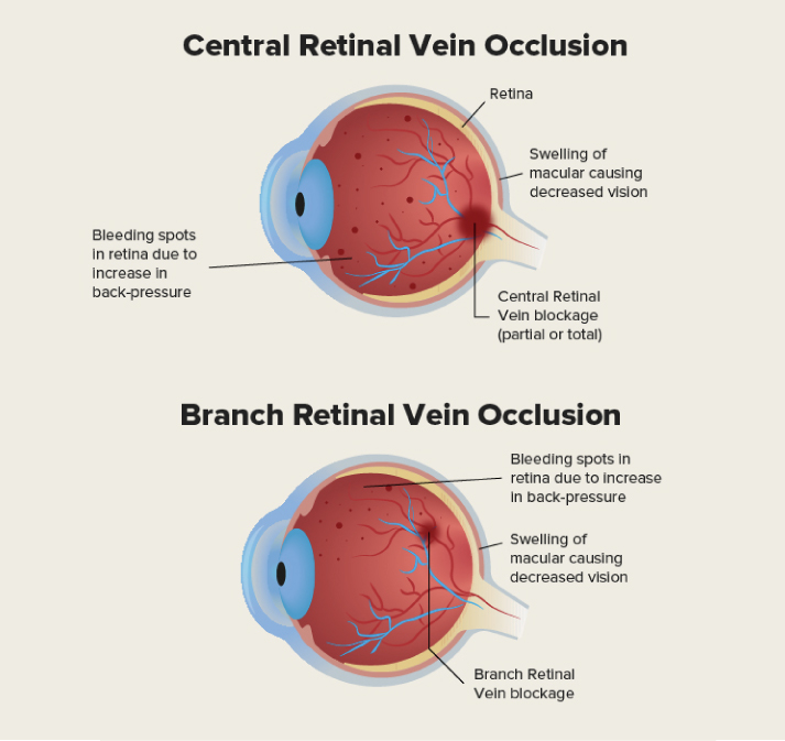
Vein occlusions block blood draining away, causing swelling and bleeding in the retina.
- Branch Retinal Vein Occlusion (BRVO) blocks a branch vein. This is the most common form.
- Central Retinal Vein Occlusion (CRVO) affects the central retinal vein and can cause more extensive retinal involvement than a branch retinal vein occlusion. CRVO can be classified as non-ischaemic or ischaemic, with ischaemic CRVO generally carrying a poorer visual prognosis and higher risk of neovascular complications.
Symptoms of a Retinal Vascular Occlusion
Retinal vascular occlusions can cause sudden, painless changes in vision. Retinal artery occlusion in particular is an ophthalmic emergency because interruption of blood flow can cause irreversible retinal damage rapidly. Sudden vision loss in one eye requires immediate medical assessment.
These are the symptoms:
- Sudden, painless blurred or reduced vision: A rapid change in vision, usually affecting one eye.
- Partial or complete vision loss: Loss of vision in part or all of the affected eye, depending on the location and extent of the occlusion.
- Distorted or wavy vision: This can occur particularly when retinal swelling affects the macula.
- Visual field changes: A retinal artery or branch vein occlusion may cause a missing or blurred area in part of the visual field.
Sudden visual loss requires immediate clinical evaluation. If you experience sudden loss or a significant change in vision, seek emergency care right away, as retinal artery occlusion can cause irreversible damage rapidly.
Noticing a change in your vision?
If you’ve been experiencing any of these symptoms, it’s important to get a thorough eye examination done. An early diagnosis and timely treatment plan from an eye specialist can give you the best chance of preserving your vision.
Book An AppointmentWho Is at Risk for Retinal Vascular Occlusion?
Several conditions that affect blood vessels and circulation can increase the risk of retinal vascular occlusion. Risk factors vary depending on whether an artery or vein is affected.
Many of these are also associated with vascular disease elsewhere in the body, which is why identifying and managing underlying risk factors is an important part of care.
Common risk factors include:
- High blood pressure
- Diabetes
- High cholesterol
- Cardiovascular disease and atherosclerosis
- Glaucoma, particularly for retinal vein occlusion
- Smoking
- Obesity
- Certain blood-clotting disorders
What Causes Retinal Vascular Occlusion?
Retinal vascular occlusion happens when normal blood flow through the retinal vessels is disrupted. The cause depends on whether a vein or an artery is affected.
In retinal vein occlusion, the blockage is associated with changes in the retinal blood vessels that can cause slowed blood flow, vessel compression and thrombus formation. In branch retinal vein occlusion, this commonly occurs where a retinal artery and vein cross, as age-related arterial changes can compress the vein and contribute to clot formation.
In retinal artery occlusion, the blockage may be caused by an embolus, such as cholesterol, platelet or other material travelling from elsewhere in the body. Common potential sources include the carotid arteries and the heart, although other causes such as inflammation, trauma or vascular spasm can also occur.
Experiencing symptoms of a retinal vascular occlusion?
See an eye specialist todaySee an eye specialist today
Are you experiencing sudden, painless blurred vision, floaters, wavy vision or partial loss of vision?
These may indicate that you have a retinal vascular occlusion and require urgent evaluation by an eye specialist.
Book an appointment todayDiagnosing Retinal Vascular Occlusion
Diagnosis begins with an examination of the eye, followed by tests to confirm the blockage and assess how much of the retina is affected. Your doctor will then look into why the blockage occurred, since the cause often lies outside the eye.
Initial checks usually include these:
- Visual acuity, tested with an eye chart
- A visual field exam, which tests your side vision
- Checking how your pupil responds to light
- Intraocular pressure, which measures the pressure in your eye
- A slit lamp exam, which lets the ophthalmologist examine the structures of your eye in detail
Imaging tests give a clearer picture of the retina itself:
- Retinal photography
- Optical coherence tomography (OCT), which uses light waves to create images of the eye
- Fluorescein angiography, which involves injecting dye into the arm and taking photos of the retina to observe how the dye circulates
Because retinal vascular occlusions can be associated with cardiovascular and systemic conditions, further evaluation may be arranged to identify underlying risk factors or, in the case of retinal artery occlusion, a possible source of emboli.
These can include blood pressure and cholesterol checks, blood tests, and scans of the neck or heart to look for a source of clots.
How Is Retinal Vascular Occlusion Treated?
Treatment depends on whether the occlusion affects a retinal vein or artery. For retinal vein occlusion, treatment focuses primarily on managing complications such as macular oedema and retinal neovascularisation.
For retinal artery occlusion, immediate medical assessment is required because irreversible retinal damage can occur rapidly. Urgent systemic evaluation is also important to identify potential causes and reduce the risk of subsequent vascular events.
Treatment for Retinal Vein Occlusion
Treatment focuses on reducing swelling, preventing abnormal vessel growth, and managing the health conditions behind the blockage.
- Anti-VEGF injections: Medications such as aflibercept, bevacizumab, or ranibizumab may be injected into the eye to treat macular oedema and reduce the effects of abnormal blood vessel growth.
- Steroid treatment: Intravitreal corticosteroids may be considered for macular oedema in selected patients. They can increase the risk of raised intraocular pressure and cataract formation, so monitoring is required.
- Laser therapy: Laser photocoagulation may be used when retinal neovascularisation develops. Depending on the type and location of the occlusion, other forms of retinal laser treatment may also be considered in selected cases.
- Vitrectomy surgery: In selected cases, vitrectomy may be considered for complications such as non-clearing vitreous haemorrhage or tractional retinal complications.
- Managing Underlying Conditions: Controlling blood pressure, blood sugar, and cholesterol supports eye health and limits future blockages.
Treatment for Retinal Artery Occlusion
Retinal artery occlusion or RAO is an emergency requiring immediate assessment. The priority is urgent evaluation for the cause of the occlusion and assessment of the patient's risk of stroke and other vascular events.
There is currently no acute treatment that has consistently demonstrated improved visual outcomes, although selected patients may be considered for treatments such as hyperbaric oxygen depending on the circumstances and local availability.
- Emergency assessment
Retinal artery occlusion requires immediate assessment, ideally through an emergency or stroke service. The priority is rapid evaluation for the cause of the occlusion and assessment of the patient's risk of stroke and other vascular events.
- Acute treatments
Historically, treatments such as ocular massage, lowering intraocular pressure and breathing carbogen have been used in an attempt to restore retinal blood flow.
However, these approaches have not consistently demonstrated a visual benefit in clinical studies. Hyperbaric oxygen may be considered in selected patients and settings, but evidence and availability vary.
- Investigating the source
Patients should undergo urgent systemic evaluation to identify potential sources of emboli or vascular disease. This may include assessment of the carotid arteries, heart, blood pressure and other cardiovascular risk factors.
Complications of Retinal Vascular Occlusion
Vision loss or visual field defects are the main consequences of retinal vascular occlusion. The impact depends on which vessel is affected and whether the macula is involved. When one eye is significantly affected, some patients may experience difficulty with tasks requiring binocular vision or depth perception.
Other complications can develop inside the eye:
- Macular oedema: Damage to retinal blood vessels can cause fluid to leak into the macula, resulting in swelling and blurred or distorted central vision.
- Vitreous haemorrhage: This is the presence of blood in the vitreous portion (clear part) of the eye.
- Neovascular glaucoma: Severe retinal ischaemia can stimulate abnormal blood vessel growth in parts of the eye, including the iris and drainage angle. These vessels can interfere with fluid drainage and cause a rise in eye pressure.
Artery occlusions carry a different risk. Because retinal tissue is highly sensitive to a lack of oxygen, prolonged interruption of its blood supply can cause permanent damage and lasting vision loss.



